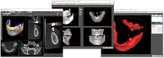
Diagnosis, from Panoramic images to CT simulation
Capturing the 3-dimensional configuration of the jawbones, which has not been available in panoramic images, to realize the diagnosis in high quality as well as in good safety.
*The LANDmarker does not include any automatic diagnosis function by the software itself or any function to design and create surgical guides.
Direct CT data conversion: Intuitive and easy CT data editing
CT data(DICOM) can be directly loaded and edited on your own computer without ordering to iLAND.
Intuitive and easy to use.
Virtual occludator
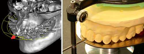
Even if the occlusal plane is tilted at the time of CT scanning, the inclination of the CT data can be corrected with the image of a dental model mounted on an articulator.
Removing metal artifacts
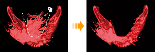
Easily and effortlessly remove unwanted metal artifacts by simply surrounding them on the 3D image.
Data creation service (charged): More accurate diagnosis
Selectable from 4 menus according to your needs
You can choose according to your needs, such as "I want to see the final prosthetic form on the data", "I want to reduce the hassle of positioning implants from scratch.", "I want to diagnose quickly with model synthesized data.".
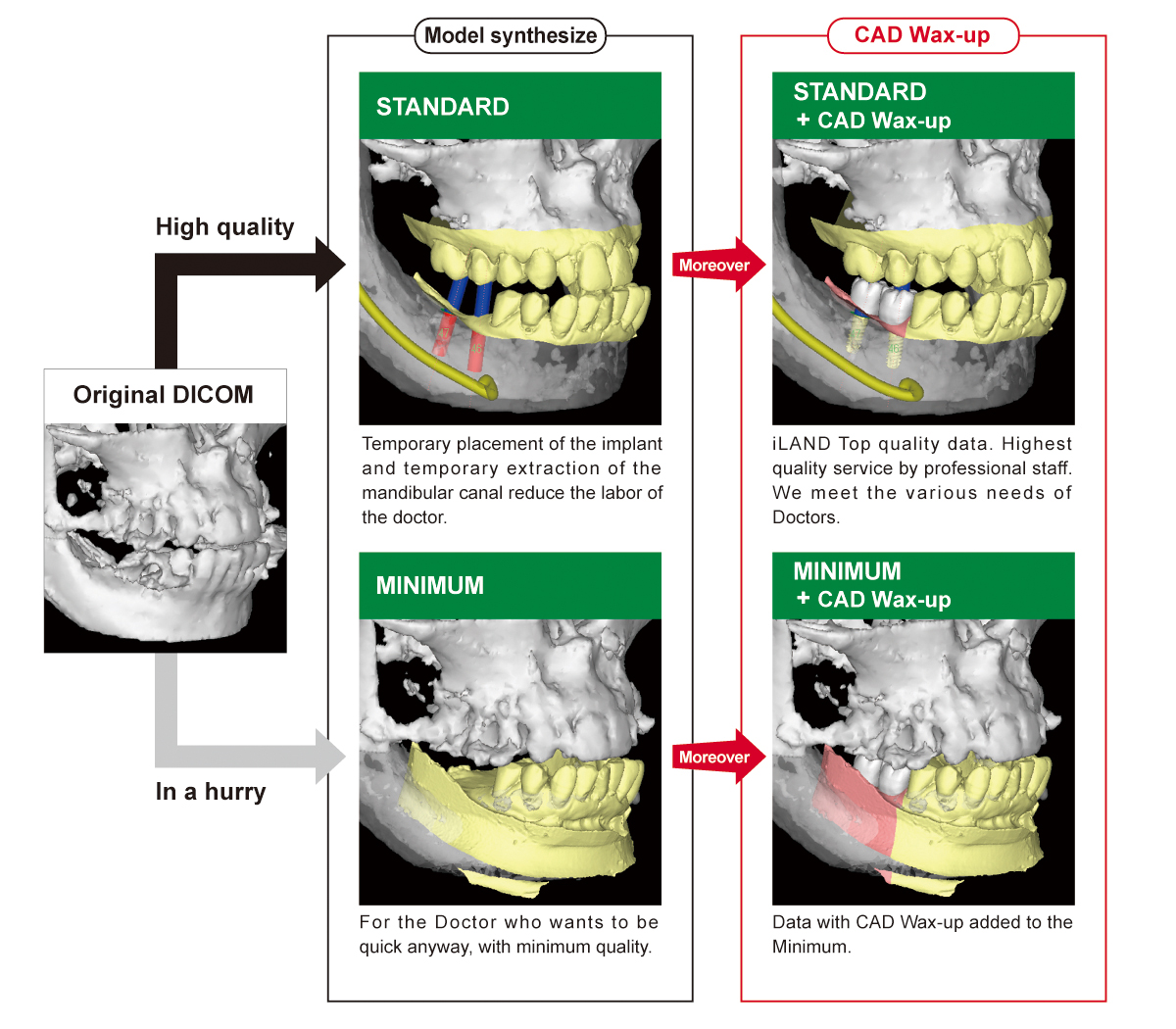
Data processing options (depending on the plan)

Clear 3D images with no metal artifacts
Create clear 3D images without metal artifacts by synthesizing CT data and study models. It is also effective for presentations to patients.
CAD Wax-up
CAD wax-up by skilled technicians is displayed on LANDmarker. This enables highly accurate top-down simulation. In addition, this CAD data can be used for provisional and final prosthesis to achieve the prosthesis as simulated.
Image of final prosthesis
The outline of the crown is displayed by synthesizing a study model with wax-up. Achieves diagnosis by top-down treatment.
Reduce the labor of Doctors
Temporary placement of the implant and temporary extraction of the mandibular canal reduce the labor of the doctor. You can diagnose as soon as you open the data.
Direct+:Data synthesis is possible at your own clinic
LANDmarker Direct+ makes it possible to perform tasks that were previously only possible with iLAND, such as "removing metal artifacts from 3D images" or "importing wax-up into simulation data for diagnosis.
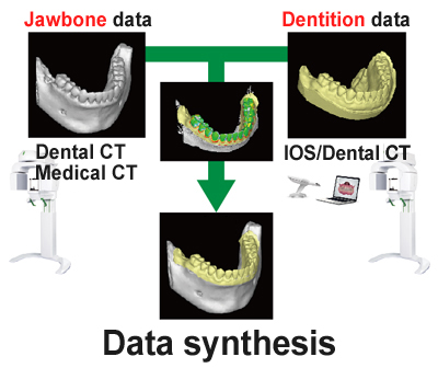
Data synthesis at your own clinic
Synthesis jawbone CT data with dentition data.
The dentition data is compatible with desktop scanners, intraoral scanners (IOS), and data of study models taken with CT.
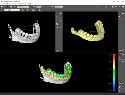
Easy synthesis by selecting 3 points
Automatic synthesis by simply selecting 3 points of the same part of CT data and model data while looking at the screen.

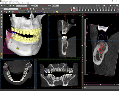
Highly accurate diagnosis, reduced cost and time
No need to send models to iLAND for synthesis, saving time and money. Trimming and model division as you like. It provides accurate diagnosis with top-down treatment and easy-to-understand patient explanation with 3D images without metal artifacts.
Multi-functional implant simulation
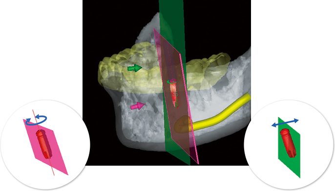
It is equipped with various functions devised by implant clinicians, including the world's first "implant cross-section" made possible by iLAND This allows for more accurate implant simulation.
Implant cross-section
 |
|
|---|---|
 |
 |
|
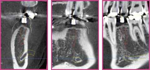
Implant cross section The cross-section along the long axis of the implant allows for accurate positioning. By rotating the cross-section, the situation around the implant can be seen at a glance. |
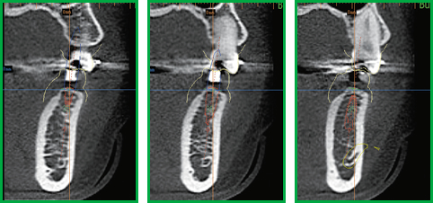
Conventional cross section perpendicular to the dental arch In the case of inclined placement, the implant is cut at an angle, making it difficult to ascertain the exact position of the implant. |
Realistic implant display
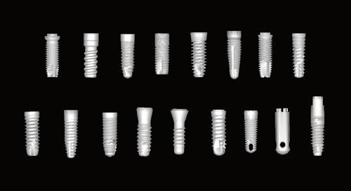
Equipped with a realistic shape with the cooperation of implant manufacturers. Simulation that is conscious of bone level is possible.
Virtual wax-up
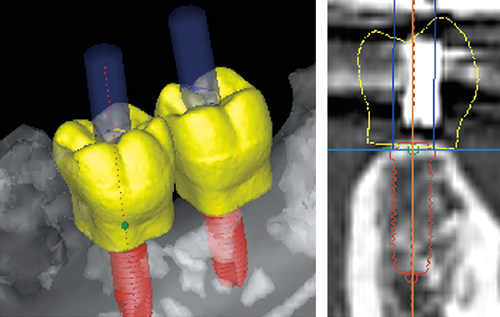
The world's first "Virtual Wax-Up" equipped with iLAND simulates the image of the final prosthesis without performing any technical work.
Implant memory
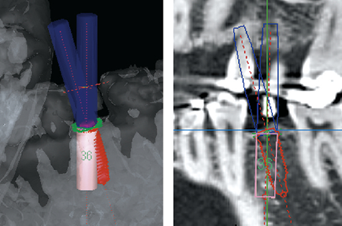
Multiple implant locations can be compared to determine a more appropriate implant location.
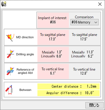
Displays the tilt angle of the implant and the distance and angle difference between implants.
Bone graft simulation
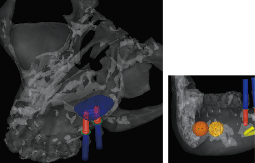
With sinus lifts or Guided Bone Regeneration, it is possible not only to measure the volume required for bone grafting, but also to measure the volume that can be collected from bones such as chins and rams.
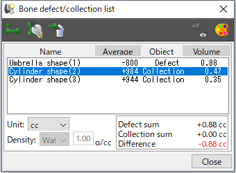
Mandibular canal marking
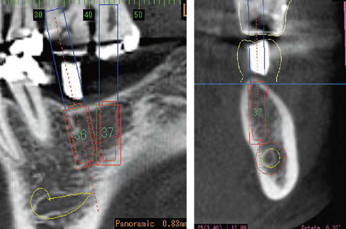
Alert display of implant-to-implant and implant-mandibular canal contact. The detection range can be set freely.

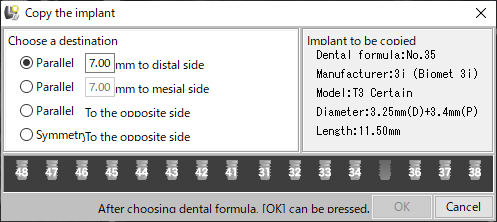
Parallel implantation
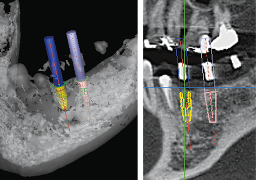
Equipped with a copy function for easy parallel implantation in mesiodistal and symmetrical implantation in the opposite side.
Other features
It is equipped with not only implant simulation but also a full range of functions. It supports the diagnosis of doctors with advanced functions.
Measurement function
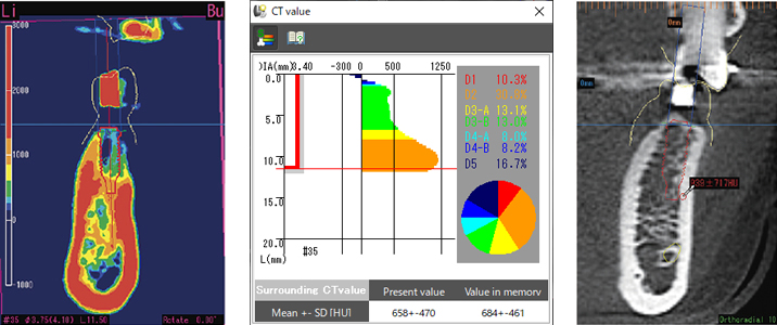
Bone quality diagnosis with CT value color display or CT value measurement.
*Please note that some dental CT models do not output CT values.
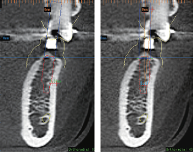
Distance and angle can be measured with tools such as a folding tape.
Automatic cross-sectional image flipping
The image is automatically inverted in the occlusal plane and in the mesial.
Prevents diagnostic mistakes by displaying images that emphasize intuition as if you were looking directly into the oral cavity.
Axial cross section
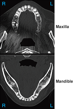
Orthoradial cross section
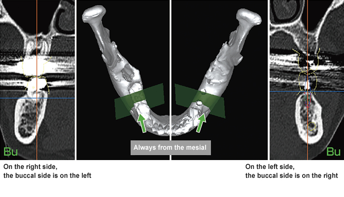
Image filter
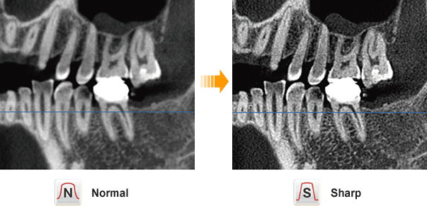
The sharpness of the image can be adjusted with the image filter function.
Ideal for informed consent
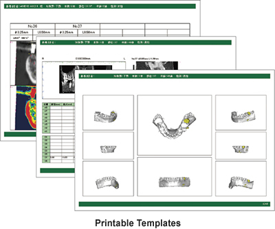
LANDmarker is also effective for informed consent. 3D image display and printing of various images are ideal for deepening the understanding of patients.
Impacted supernumerary teeth
3D images are displayed as surface rendering or volume rendering. Patients are satisfied with the easy-to-understand images.
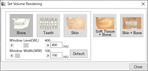
In volume rendering, the extraction range can be changed by switching buttons.
*For dental CT that does not output CT values, it is necessary to adjust the extraction range.

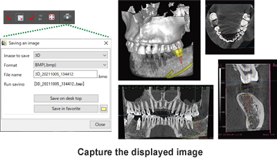
Printing and camera function
